Activity
Mon
Wed
Fri
Sun
Oct
Nov
Dec
Jan
Feb
Mar
Apr
May
Jun
Jul
Aug
Sep
What is this?
Less
More
Memberships
MSK Radiology
2k members • Free
Virtual MSK Fellowship
384 members • Paid
38 contributions to MSK Radiology


1 like • Jul 8
ALT? Needs more workup like biopsy - let us know, what happens next!

Jul 8 •
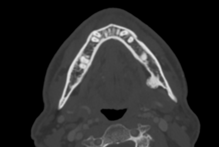
Bony Mandible lesion
Male 58 y.o. Referred due to a long standing painless left hemimandible lesion. Solid and inmovile to palpation. Somebody did an US (I don't have images nor report) and send to CT (soft tissue window unremarkable, I've only uploaded bone window). Bony lesion, left hemimandible. Cortical continuity without a worrisome cleft. I'm leaning towards osteoid osteoma due to frequency and location, but I'm doubting if it presents a calcified thin layer of cartilaginous cap. Would you recommend further workup? Differential Turret exostosis / NORA, anything else? https://www.cmrad.com/cases/1834740247
1 like • Jul 8
Parosteal Osteoma and Osteochondroma comes to mind. cor and axial MRI (plain 2D T2, 2D T1 and maybe fs PD) images will help to find a possible cartilage layer. Let us know what happens next!

Jun 21 •
Guidance while starting out
Hi, I am a radiology resident wanting to get started with msk and want to build up good foundation. Could you please guide me with how shall I start ?
3 likes • Jun 23
I'm totally with Im Beg: - (MR-)anatomy is key - Go joint by joint, start with a frequently reported one like the knee, don't start with lumbosacral plexus MRI ;) - I would rather stick to online resources than books. - When you reached rheumathologic diseases, there is no way getting around Prof. Hermann and the Berlin Case viewer - If you are technophil go for the everythingMRI youtube-channel BW, Max
Jun 19 •
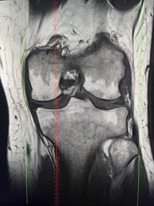
Bone marrow
There is a case of 45 yo female. The examination was done due to injury in 15th of March. I have a question about bone marrow reconvertion. I can see T1 low signal involved partially subchondral layers of tibial and femoral condyles. Any suggestions about pathologic process?
3 likes • Jun 20
I'm with Im Beg, because the signal is still higher than the muscle matrix. Looks familiar in this article: (PDF) Common Mistakes and Pitfalls in Magnetic Resonance Imaging of the Knee Reconversion of the bone marrow from yellow to red
Jun 17 •
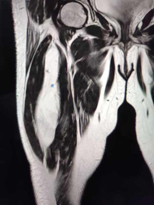
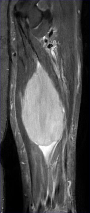
Primary and secondary (muscle) lymphoma
Hi guys, the first case I discussed a year ago with @Esteban Peghini in one of the Q&A Sessions within the fellowship. He straight foreward said: Could be lymphoma. We came up with some DD's as well and recommended biopsy. Since this was just a single case I didn't do a FU and didn't got my handy on the histology report. Today I had a similar case again and was curious what happened to my patient last year - so I talked to the surgeon asking for the histology report. First patient was a confirmed case of diffuse large B-cell lymphoma. The patient from today turned out to have a known and currently treated mantle cell lymphoma. Since the rapidly progrentient swelling of the forearm in case 2 is develloping fast there will be a biopsy as well. Firstly to excluse a large median nerv neuroma and second to compare the cell clones in case of a mantle cell lymphoma of the forearm resistent to systemic therapy. case 1: confirmed DLBCL DLBCL - diffuse large B-cell lymphoma - Collective Minds Radiology case 2: suspected case of mantle cell lymphoma, DD neurinoma mantle cell lymphoma - Collective Minds Radiology I hope you'll find these cases interesting as well and best wishes, Max
1-10 of 38

Active 4d ago
Joined Jan 15, 2023
Germany
Powered by