Activity
Mon
Wed
Fri
Sun
Nov
Dec
Jan
Feb
Mar
Apr
May
Jun
Jul
Aug
Sep
What is this?
Less
More
Memberships
MSK Radiology
2k members • Free
RS
Radiology School
52 members • $5/m
8 contributions to MSK Radiology
Aug 13 •
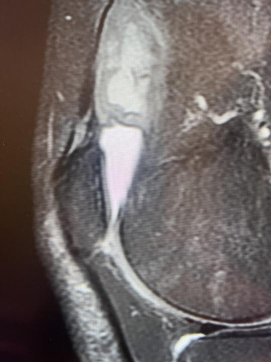
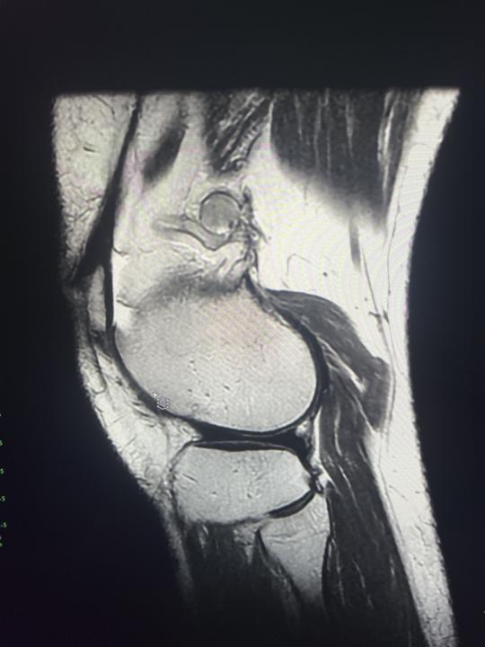
Suprapatellar lesion
35 yo male with vague knee pain since 2 weeks. There is a suprapatellar lesion. Do you have an idea what could it be? Fluid with septa and thickened synovium? Hematoma? PDfs, T1W and T2W images are obtained. Additionally partially torn ACL was depicted.
0 likes • Aug 13
Thanks
Jul 31 •
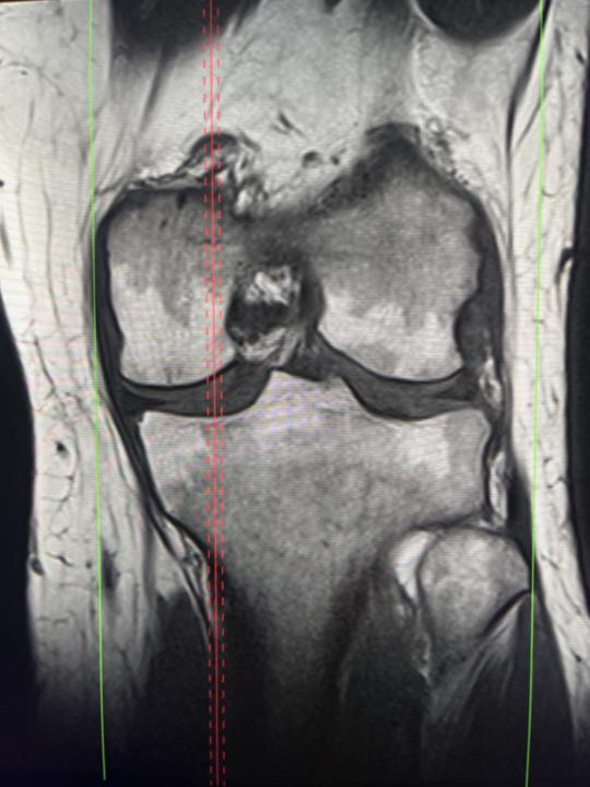
Knee pain
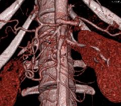
57 yo female with knee pain. Baker cyst was suspected by referring physician. I can see small amount of fluid in SB and partial cartilage loss on MFC. On the lateral side of femoral bone I suspect thrombosis of varicose vein. Can you agree or do you have another suggestions?
0 likes • Aug 3
Thanks
Jun 19 •
Bone marrow
There is a case of 45 yo female. The examination was done due to injury in 15th of March. I have a question about bone marrow reconvertion. I can see T1 low signal involved partially subchondral layers of tibial and femoral condyles. Any suggestions about pathologic process?
0 likes • Jun 20
Thank you
Dec '24 •
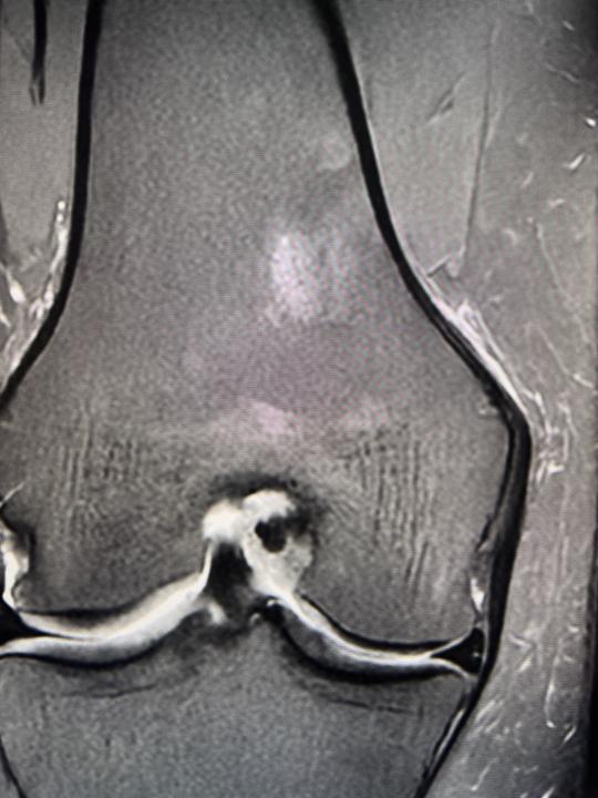
Focal area in distal femoral bone
There is a focal area of high signal in PD FS, slightly decreased in T1, T2 and T2 GRE. It is localized in posterior part of distal femur and has not distinct borders, no changes in cortical part of bone. Would you include it in you report and recommend further investinagion in x-ray and MR with contrast? Patient is a 44 yo female with unspecific symptoms. Photos: PD FS, T2, T2 GRE, T1, T2 GRE
0 likes • Dec '24
Thanks
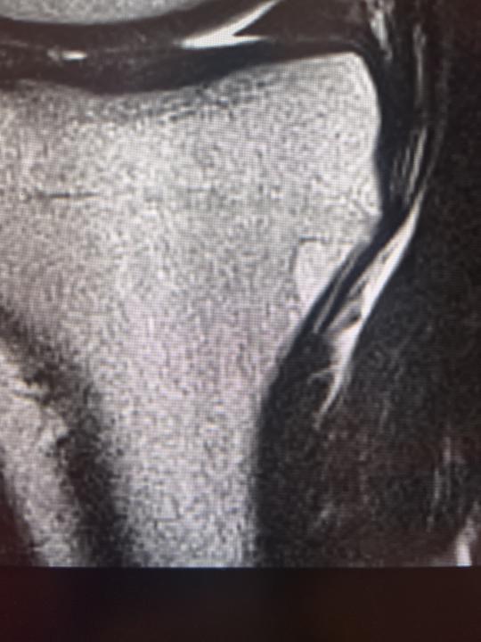
Nov '24 •
Proximal tibial lesion
There is a lesion in proxial tibia in 35yo male. It has homogenous T1 and T2 high signal and PD fs low signal. Do you think it may be an intraosseus lipoma? Photos: T2, PD fs, T1.
1-8 of 8
