Activity
Mon
Wed
Fri
Sun
Nov
Dec
Jan
Feb
Mar
Apr
May
Jun
Jul
Aug
Sep
Oct
What is this?
Less
More
Memberships
MSK Radiology
2k members • Free
34 contributions to MSK Radiology

Sep 1 •
The Agten Library
MSK Radiology You Won't Find in Textbooks. I have been working over the last couple of months on this project. It is a curated collection of over 1260 short videos in which I answer practical questions of fellows from the Virtual MSK Fellowship. https://library.agten.org

0 likes • Sep 6
Good job Christoph! Like the Lucas arts archives style. Does 7 day trial include everything in the library?
Aug 25 •
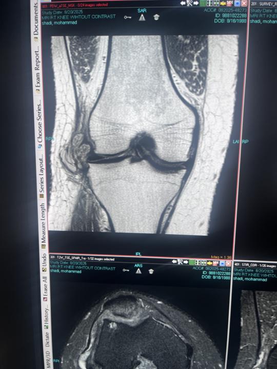
Knee MRI
Hello Please I need your help in this case No post contrast image ,history of knee scope 3yrs ago
0 likes • Sep 6
Seems to be big parameniscal ganglion. Have some junior general practiotioner put needle in to it before MRI?

Aug 25 •
Humeral anchor dislodgement / migration of long head of biceps tenodesis ?
Is there medial migration of the tenodesis screw? I seem to see a T2 hyperintense signal at the location of the screw, with the metal artifact displaced medially and posteriorly, and extending beyond the posterior cortex of the humerus.

1 like • Sep 6
Seems to be normal finding.

2 likes • Sep 6
Yeah with quick glance it's extensive partial cuff rupture with perforation overlap area and bursal surface delamination of SSc. LHB tendon is extensively partially ruptured and luxated.
Aug 29 •
Thigh pain
Woman 51. Referred due to mid thigh pain after fall. She says the pain has been there for years already, worsened with the fall. CT and MRI are 10 days apart. No history of previous illness but carpal tunnel. No relevant medication. My doubt is differentiating Osteoid Osteoma from old stress injury. There is BME and cortical seems a little bit irregular. Course of action? If O.O. confirmed I can perform cryoablation and treat it. Doubting this is a nidus. Thanks in advance! https://www.cmrad.com/cases/1765544550
1 like • Sep 6
Hard to say, not typical age for osteoid osteoma though. There is some cracking of the periosteal cortex also though that navigates the diagnostic toward chronic stress fracture. Some sort of metabolic imaging eg. scintigraphy would do the trick just like Dr Beg suggested.
1-10 of 34

Active 1d ago
Joined Apr 4, 2025
Powered by