Activity
Mon
Wed
Fri
Sun
Nov
Dec
Jan
Feb
Mar
Apr
May
Jun
Jul
Aug
Sep
Oct
What is this?
Less
More
Memberships
MSK Radiology
2k members • Free
20 contributions to MSK Radiology

Apr 18 •
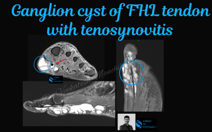
Large Ganglion Cyst of the Flexor Hallucis Longus (FHL) Tendon
🧠 Teaching Case: Large Ganglion Cyst of the Flexor Hallucis Longus (FHL) Tendon with Tenosynovitis on MRI 📌 Clinical Context: A 60-year-old patient presents with pain along the foot. No history of acute trauma. Pain is exacerbated by plantarflexion, especially during activities 📸 MRI Findings: 👉 Sagittal and axial T2-weighted fat-saturated images reveal: A multiloculated, well-defined hyperintense cystic lesion(Blue circle) along the course of the FHL tendon, centered in the forefoot and mid foot region Tenosynovitis: Surrounding high signal fluid within the tendon sheath with mild tendon sheath thickening.(Red arrow) The FHL tendon is intact but slightly flattened within the tunnel, indicating mass effect from the cyst. 🧬 Diagnosis: 🔹 Ganglion cyst of the flexor hallucis longus tendon sheath 🔹 Associated tenosynovitis 🧠 Pearls for Practice: ✅ Ganglion cysts are benign mucin-filled synovial cysts, often arising from joint capsules or tendon sheaths. In the ankle, FHL is a common site due to repetitive motion and mechanical stress. ✅ Tenosynovitis may be reactive or secondary to chronic friction from the cyst. In athletes and dancers, FHL involvement is especially common due to overuse. ✅ MRI is the modality of choice: T1: Hypointense T2/STIR: Hyperintense, well-circumscribed Look for communication with joint or tendon sheath ✅ Clinical Correlation: Symptoms may mimic tarsal tunnel syndrome or posterior ankle impingement. 🩺 Management: Conservative: NSAIDs, rest, activity modification Interventional: Ultrasound-guided aspiration Surgical: Excision if symptomatic or recurrent
0 likes • Apr 21
@Tapas Sahu yes
0 likes • Apr 21
@Tapas Sahu I have edited that
Mar 13 •
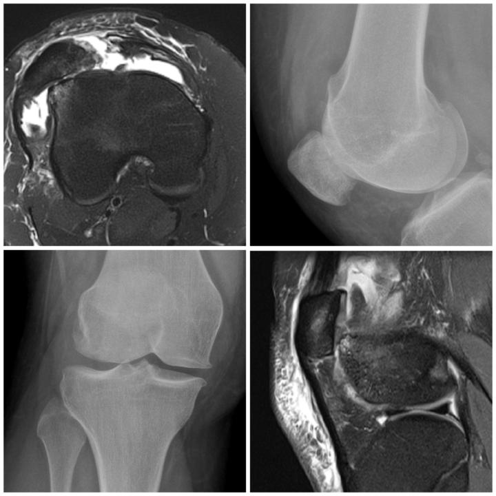
Trochlear dysplasia with MPFL tear
✅✅"Case Spotlight: Trochlear Dysplasia with Patellar Dislocation and MPFL Tear on MRI As a radiologist, I'd like to share a complex case involving trochlear dysplasia, patellar dislocation, and a tear of the medial patellofemoral ligament (MPFL). Clinical Presentation:🌟 - Recurrent patellar dislocations - Knee pain and instability MRI Findings:✅ - Trochlear dysplasia with a shallow femoral trochlea - Patellar dislocation with lateral patellar tilt - Complete tear of the MPFL - Associated bone marrow edema and soft tissue injuries Trochlear dysplasia is a critical factor in patellar instability, and MPFL tears are a common consequence of patellar dislocation. Key takeaways:⚜️⚜️ - MRI is essential for diagnosing trochlear dysplasia and MPFL tears - Accurate diagnosis guides surgical planning and reconstruction - MPFL reconstruction is often necessary to restore patellar stability✅
2
0
Mar 8 •
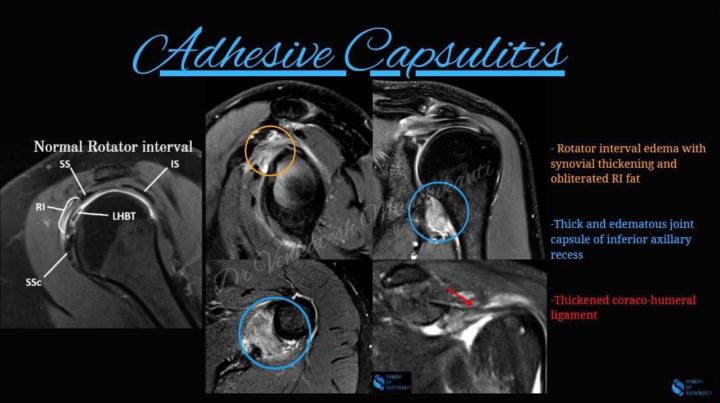
Adhesive Capsulitis
✅"Case Spotlight: Adhesive Capsulitis on MRI🌟 As a radiologist, I'd like to share a fascinating case of adhesive capsulitis, also known as frozen shoulder. Clinical Presentation:⚜️ - Gradual onset of shoulder pain and stiffness - Limited range of motion and difficulty with daily activities MRI Findings:🌟 - Thickening and enhancement of the joint capsule - Adhesions and fibrosis within the capsule - Possible reduction in joint volume and effusion Adhesive capsulitis can be challenging to diagnose and treat. MRI plays a crucial role in confirming the diagnosis and guiding management. Key takeaways:🦾🦾 - MRI is essential for diagnosing adhesive capsulitis - Joint capsule thickening and enhancement are hallmark findings - Early diagnosis and treatment can improve outcomes
0 likes • Mar 13
@Vikrant Pulliwar Pls click on the image Dr you can see the description of coloured annotations
Mar 6 •
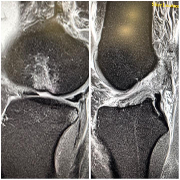
Lateral Femoral Sulcus Sign
"ACL Tear Alert: Lateral Femoral Sulcus Sign on MRI🌟 As a radiologist, I'd like to highlight a crucial MRI finding that can indicate an ACL tear: the Lateral Femoral Sulcus (LFS) sign.🌟 The LFS sign refers to a focal depression or sulcus in the lateral femoral condyle, which can be seen on sagittal MRI images. This sign is highly specific for ACL tears, particularly in the acute setting.⚜️ Why is the LFS sign important?🦾 - High sensitivity and specificity for ACL tears - Can be seen in the absence of other obvious ACL injury findings - May indicate a more severe ACL injury When interpreting knee MRI scans, don't miss this subtle but critical sign!🌟
Mar 6 •
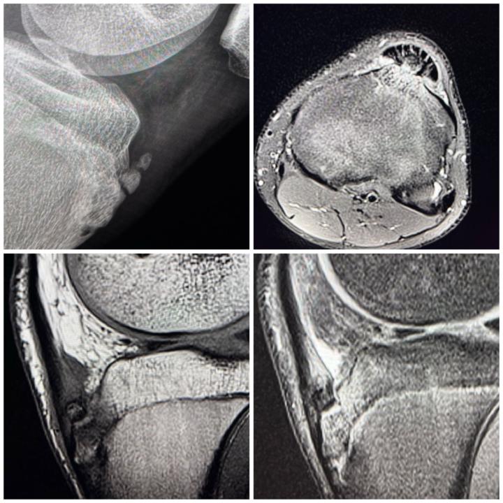
⚜️Osgood-Schlatter disease
🌟"Case Spotlight: Osgood-Schlatter Disease on X-ray and MRI⚜️ As a radiologist, I'd like to share a fascinating case of Osgood-Schlatter disease, a common cause of knee pain in adolescents. Clinical Presentation: - Pain and swelling below the kneecap - Typically affects active teenagers during growth spurts Imaging Findings: X-ray:🌟 - Soft tissue swelling and fragmentation of the tibial tuberosity MRI:🌟 - Edema and inflammation of the patellar tendon and surrounding soft tissues - Fragmentation and heterogeneity of the tibial tuberosity ⚜️Osgood-Schlatter disease is a self-limiting condition that resolves with maturity. However, imaging plays a crucial role in confirming the diagnosis and ruling out other potential causes of knee pain.
3
0
1-10 of 20

Active 77d ago
Joined Jan 6, 2025
United Arab Emirates
Powered by