Activity
Mon
Wed
Fri
Sun
Sep
Oct
Nov
Dec
Jan
Feb
Mar
Apr
May
Jun
Jul
Aug
What is this?
Less
More
Owned by Nabila
Cardiology education & community for medical students, nurses, PAs & doctors – clear, practical, career-focused.
31 contributions to The Cardiology Tutor
3d •
Title: The biggest cardiology myth I was taught that turned out to be wrong 🫀
I want to do something different today. I have been a Cardiology Consultant for a few years. I trained at some of the best institutions in the country. And in that time, I was taught things about cardiology that turned out to be either wrong, incomplete, or dangerously oversimplified. I will start. The myth I was taught: “A normal ECG rules out a heart attack.” I heard this repeatedly. In lectures. On ward rounds. In revision guides. It is not true. Up to 20% of STEMIs can present with a completely normal or near-normal initial ECG. Posterior STEMIs, in particular, are notorious for this, the changes are there, but they are subtle and easily missed if you are looking for the classic ST elevation pattern you were taught. The first ECG is a snapshot. A single moment in time. Serial ECGs, troponin trends, and clinical context are what actually tell the story. A normal ECG in a patient with typical cardiac chest pain should never be the reason to stop looking. I have seen that myth cause harm. I have seen it almost cause harm to my own patients. And that is why I teach it differently now. Your turn. What is the biggest cardiology myth - or clinical teaching - you were given that turned out to be wrong, incomplete, or that you wish someone had corrected earlier? No wrong answers. No judgment. This is exactly the kind of conversation that makes good doctors and nurses better clinicians. Drop it below. 👇 —
0
0
11d •
ECG Case #1 - Monday Morning on the Take 🫀
Good afternoon everyone. Starting today, I’m posting a new clinical case every week in this community. No slides, no lecture format - just a real scenario, an ECG, and a question. The format Sumbel asked for. If it works for you too, tell me below and I’ll make it daily. Here’s Case #1. The scenario: You’re the FY1 on call. It’s 8am Monday. You’re called to see a 67-year-old woman on the medical ward. She was admitted yesterday with two days of palpitations and mild breathlessness. Otherwise stable. No chest pain. Background of hypertension and type 2 diabetes. Her observations this morning: HR 138, BP 102/68, O2 sats 96% on air, RR 18, afebrile. The nurse hands you this morning’s ECG. ECG findings: • No discernible P waves • Irregularly irregular ventricular rhythm • Ventricular rate approximately 138 bpm • Narrow QRS complexes throughout • No ST changes Three questions for you. Answer in the comments: 1. What’s the diagnosis? 2. She’s been in this rhythm for over 48 hours. What’s the single most important thing you need to establish before cardioverting her? 3. Her BP is 102/68. Does that change your management? If so, how? No wrong answers here. This is a safe space to think out loud. I’ll post the full discussion and teaching points in 24 hours - including the one thing most junior doctors miss in this exact scenario. If you want to tag a colleague who would benefit from working through this, do it below. The more people in the room, the better the discussion. Drop your answers 👇
11d •
Need your input
👋 Welcome - and I want to hear from you First things first. If you're reading this, you're already part of something I'm genuinely proud of. This community is free right now. And I want to keep giving you real, consultant-level cardiology education - no paywalls, no watered-down content - for as long as I possibly can before we build out the full membership. But I need your help to make this worth your time. So I'm going to ask you three questions. Answer one, answer all three, or just introduce yourself below. Every response helps me build this community around what you actually need. Question 1 — Who are you? Drop your role and where you are in your training. Medical student? FY1? Nurse? Paramedic? Pharmacist? Cardiology registrar? Something else entirely? I want to know exactly who's in this room. Question 2 — What's the ONE cardiology topic that makes you feel underprepared? Be honest. There's no wrong answer here and nothing is too basic. The most common answers in the last community I asked this were: - ECG interpretation under pressure - Managing AF on the acute take - When to worry about a troponin result - What to say when you call the cardiology registrar - Echocardiography — what it shows and when to request it What's yours? Question 3 — What would make this community genuinely useful to you? Daily ECG cases? Weekly live Q&As? Case-based discussions? A structured learning pathway? A place to ask clinical questions without judgment? Tell me what would make you come back every day. Here's what I'm committing to in return: ✅ Weekly ECG cases with full consultant interpretation - posted every Monday ✅ Monthly live cardiology teaching sessions - free for all members ✅ A resource library building up over the coming weeks - ECG guides, clinical frameworks, drug references ✅ Direct access to me to ask clinical questions - I'll answer every one personally to start with The only thing I ask from you: show up, engage, and share this community with one colleague who would benefit.
0 likes • 11d
@Sumbel Khan Sumbel, thank you for being so honest and so precise about what you’re experiencing because what you’ve just described is one of the most clinically intelligent observations I hear, and most people don’t have the self-awareness to articulate it the way you did. The “excluding rather than including” feeling with ECGs is real, and it’s not a knowledge gap - it’s a pattern recognition gap. You’ve been trained to work systematically through a differential, which is exactly the right approach for most of medicine. ECG interpretation asks your brain to do something slightly different: to see a pattern before you reason through it. That shift feels uncomfortable until it clicks, and when it does, it clicks fast. You’re an IMT doctor managing real cardiology patients. The fact that you can feel that gap and name it precisely means you’re closer to bridging it than you think. And for what it’s worth - almost every IMT I’ve ever taught has stood exactly where you’re standing right now. It’s not a you problem. It’s a training gap in the system. Daily ECG cases with clinical vignettes and text on screen - that’s exactly what I’m building here, and your comment has just confirmed it’s the right thing to prioritise first. Watch this space. If anyone else in the community feels the same way Sumbel does - and I suspect most of you do - please say so below. The more you tell me where the gaps are, the more precisely I can fill them. That’s what this community is for. 🫀
13d •
Welcome (to the newbies)
If you're here, you're already ahead of most healthcare professionals - because you're actively choosing to learn outside of what's handed to you. I'm Dr Nabila, Cardiology Consultant at St Bartholomew's Hospital, London. I built this community for one reason: the gap between what medical education teaches and what clinical cardiology actually demands is too wide, and in this specialty that gap costs lives. Here's what you'll get in this community: - ECG cases with full consultant interpretation - Clinical pearls from the cardiac take - A space to ask questions without feeling judged - Advance access to everything TCT releases To start: download the free 50 ECG guide [link] — it's the reference I wish existed when I was an FY1. Introduce yourself below. What's your role, and what's the one cardiology topic that keeps you up at night? 👇
1
0
Jun 16 •
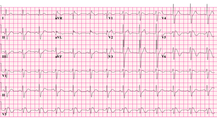
This ECG got a completely healthy 23-year-old banned from competitive sport. Why?
Question: What is the MOST likely diagnosis? Answer revealed tomorrow but I want to see your answer and reasoning. Good luck!🤞
Poll
4 members have voted
0 likes • Jun 17
🚨 ANSWER REVEALED 🚨 The diagnosis is: ✅ Brugada Syndrome Type 1 This ECG is considered diagnostic. Key clue #1 Don’t focus on the QRS. Focus on V1-V3. Key clue #2 Notice the: - Coved ST elevation - Downsloping ST segment - T-wave inversion This is the classic Type 1 Brugada pattern. Why was the athlete banned? Because Brugada Syndrome is associated with: ⚠️ Ventricular fibrillation ⚠️ Sudden cardiac death ⚠️ Syncope ⚠️ Nocturnal arrhythmias Many patients are completely healthy until their first arrhythmic event. Teaching Pearl The most dangerous ECGs are often not the ugliest. This patient could have: - Normal echocardiogram - Normal exercise capacity - No symptoms Yet still be at risk of sudden cardiac death. That’s why recognising Brugada matters.
1-10 of 31
@nabila-laskar-6144
Cardiology education & community for medical students, nurses, PAs & doctors – clear, practical, career-focused. only with Dr Nabila Laskar
Active 3d ago
Joined Sep 11, 2025
London